| Sincich, LC, Jocson, CM, Horton, JC. "Neurons in V1 Patch Columns Project to V2 Thin Stripes." Cerebral Cortex 2006; doi:10.1093/cercor/bhl004. |
Fig. 1
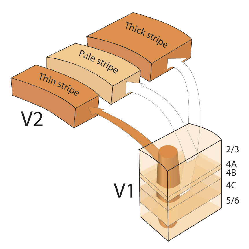
Fig. 2
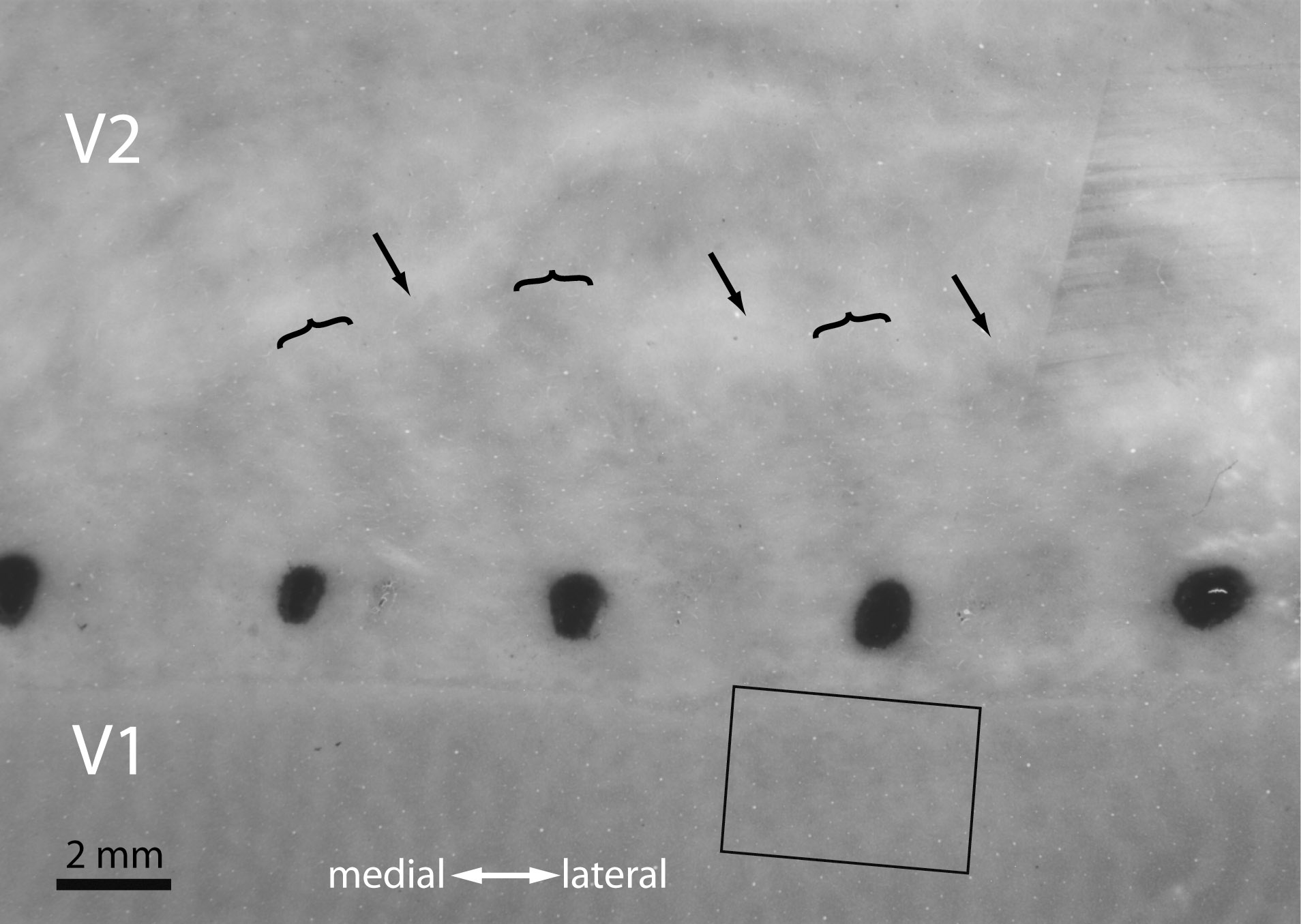
Fig. 3
Fig. 4
| Horton JC, Parker AB, Botelho JV, Duncan JL. "Spontaneous Regeneration of Human Photoreceptor Outer Segments." Sci Rep 2015; doi:10.1038/srep12364. |
Fig. 1
Fig. 2
Fig. 3
Fig.4
Fig. 5
Our research on the human visual system is based on clinical experience with patients suffering from diseases that produce eye movements abnormalities, double vision, visual field deficits, optic nerve and retinal dysfunction. Through neuro-ophthalmological examination, neuroradiological imaging, electrophysiology, pathology, and laboratory studies we characterize deficits in function to arrive at a diagnosis. This process allows us to serve patients, and to further elucidate the natural history, pathogenesis, and treatment of neuro-ophthalmological diseases.
In addition, we conduct studies of human brain tissues willed by patients to medical science. Through post-mortem examination of the occipital lobes we have been able to identify important functional elements of the human visual cortex. Figure 18 shows a montage of sections through layer 4c, processed for cytochrome oxidase, from a man who died several years after enucleation of one eye. A pattern of alternating light and dark columns are visible. The pale columns correspond to the ocular dominance columns of the missing eye:
Fig. 18) Montage of cytochrome oxidase sections through layer 4c cut through striate cortex along the medial face of the right occipital lobe, showing ocular dominance columns. They appear visible because loss of physiological activity in the...
Amblyopia is defined as a disease caused by abnormal visual experience during early childhood, which results in a decrease in acuity that can not be explained by pathology within the eye itself. For example, if a child is born with a dense cataract in one eye, the retina will be deprived of normal visual stimulation (Fig. 13). When the child grows up, the visual acuity in the affected eye will be poor -- even if the cataract is removed later by surgery and replaced by a clear lens of the appropriate refractive power. Without an ocular explanation for the permanent loss of vision caused by visual deprivation, investigators have long suspected that amblyopia is caused by anomalous wiring of the eye's central connections in the brain. This view has been confirmed by experiments suturing closed the lids of one eye in kittens or monkeys at an early age to simulate cataract (Wiesel and Hubel 1963, 1965; von Noorden et al. 1970). It is important to note that amblyopia develops only in the young, when the visual system is still immature, and hence vulnerable to the effects of deleterious environmental manipulations. This brief interval of early vulnerability is called the “critical period”.
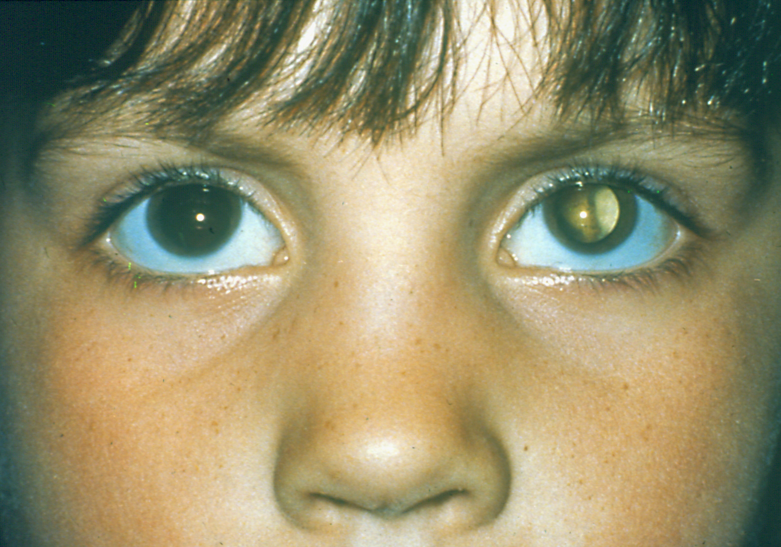
Fig. 13) A child with amblyopia in the left eye due to a congenital cataract. Prolonged deprivation from the cataract can cause rewiring of connections in the visual cortex, leading to permanent...
The cerebral cortex is a rather disappointing tissue at first glance. Its surface appears as a convoluted, gray sheet of tissue with no recognizable boundaries. In cross-section, architectonic borders can be identified with stains for cells and myelin, but only between a few areas. In a classic work, Korbinian Brodmann compiled a complete histological survey of the cerebral cortex, dividing it into 47 distinct areas. Sadly, most of the boundaries he drew have not stood the test of time.
For example, in the visual cortex Brodmann recognized three regions: areas 17, 18, and 19. Area 17, the primary visual cortex (V1), is easy to identify because it contains a prominent myelin band, the stria of Gennari, and a precise representation of the contralateral field of vision. However, the boundary between area 18 and 19 is invisible, and it is clear that there are far more than 3 visual areas in the primate brain. After processing in area 17, visual information is distributed for further analysis to at least a dozen surrounding visual areas. These areas are known collectively as “extrastriate visual cortex”. Mapping the boundaries, organization, and projections of these visual areas is a difficult challenge, in part because the primate cerebral cortex is highly folded into sulci and gyri. To facilitate anatomical studies of the macaque cortex, we have developed a method for flattening it prior to preparation of histological sections. The flatmount technique allows one to obtain single sections of the entire cortex, greatly facilitating the analysis of anatomical data. Figure 9 shows an example of a flattened hemisphere from a macaque monkey:
...In 1782 a young medical student named Francesco Gennari described a thin white stripe of myelin running through the gray matter of the occipital lobe. This stria, visible to the naked eye in fresh or fixed specimens, prompted him to suggest that the cortex might be subdivided into discrete anatomical regions. This suggestion was revolutionary, because anatomists had previously assumed that the cortex was a uniform sheet of tissue, lacking any internal subdivisions. Today, of course, it is a well accepted notion that the cerebral cortex is partitioned into dozens of distinct areas controlling all aspects of human behavior. It was Gennari's discovery which opened the door for our modern view of cortical function. Ironically, in identifying the first cortical area in the brain, Gennari had no inkling that he had stumbled upon the primary visual cortex. More than a century was to elapse before it was finally proven by Henschen, a Swedish neuropathologist, that the stria of Gennari is coextensive with the primary visual cortex. The primary visual cortex is often called the "striate cortex", recalling the prominent white stria found by Gennari.
The representation of vision in the cerebral cortex was explored early in this century by the clinical examination of soldiers wounded in battle. Inouye constructed the first detailed retinotopic map of primary visual cortex by matching visual field deficits with the trajectory of missiles penetrating the occipital lobes. We have prepared a revised version of these maps (Fig. 6) with the aid of magnetic resonance, a high-resolution imaging technique that allows direct correlation of occipital lobe lesions with visual field defects...
Strabismus is a disease characterized by misalignment of the eyes that affects 1-2% of the population of the United States. Normal stereovision is impossible, because the eyes are unable to fuse together on a target. As a result, children with strabismus have poor depth perception, impairing their ability later in life to perform certain jobs, to compete in sports, and to enjoy a normal three-dimensional view of their environment. In addition, many children with strabismus eventually develop amblyopia, depriving them of normal sight in one eye. If anything happens to their good eye, they face a grave predicament. There are also intangible, but significant, psychological aspects to strabismus. Because direct eye contact is so important in social interactions, patients with strabismus are sometimes at a disadvantage, and may suffer discrimination when seeking employment.
The crucial point is that strabismus occurs without any weakness of the eye muscles, abnormality of the cranial nerves, or intrinsic disorder of the eyes. The primary culprit is a failure of the brain mechanisms responsible for establishing or maintaining binocular fusion. Our goal is to elucidate these neural deficits. We are motivated by a conviction that understanding these deficits is essential to finding better methods of preventing and treating strabismus.
To make progress against strabismus, we employ a two-pronged approach by conducting research in both humans and monkeys (Fig. 1). Laboratory studies in humans have allowed us to define more precisely the ocular motor and perceptual abnormalities that are present in strabismus. Observations made in...
(A) Recordings from a 26-year-old woman (plano OU) during fixation on a 0.5° target at 57 cm. The mean position (solid line) and standard deviation (shading) are shown for the right eye (red) and left eye (blue). At t = 0 s, a shutter occluded either the right eye or the left eye, inducing an exodeviation. n = number of events. Positive and negative values for horizontal deviation denote right and left gaze respectively. (B) Spontaneous exotropia occurring intermittently between cover-induced episodes has an amplitude nearly equal to shutter-induced exotropia, with a difference of 0.1° for right exotropia and 0.4° for left exotropia. The shapes of the mean position traces and the variability in individual position traces are also similar. Traces are aligned at t = 0 s, with movement onset. (C) Recordings from a 36-year-old woman (-1.00 sphere OU), showing a larger exotropia. (D) Interspersed episodes of spontaneous fusion loss are similar to those from shutter occlusion, with an amplitude difference of 0.7° for right exotropia and 1.2° for left exotropia.
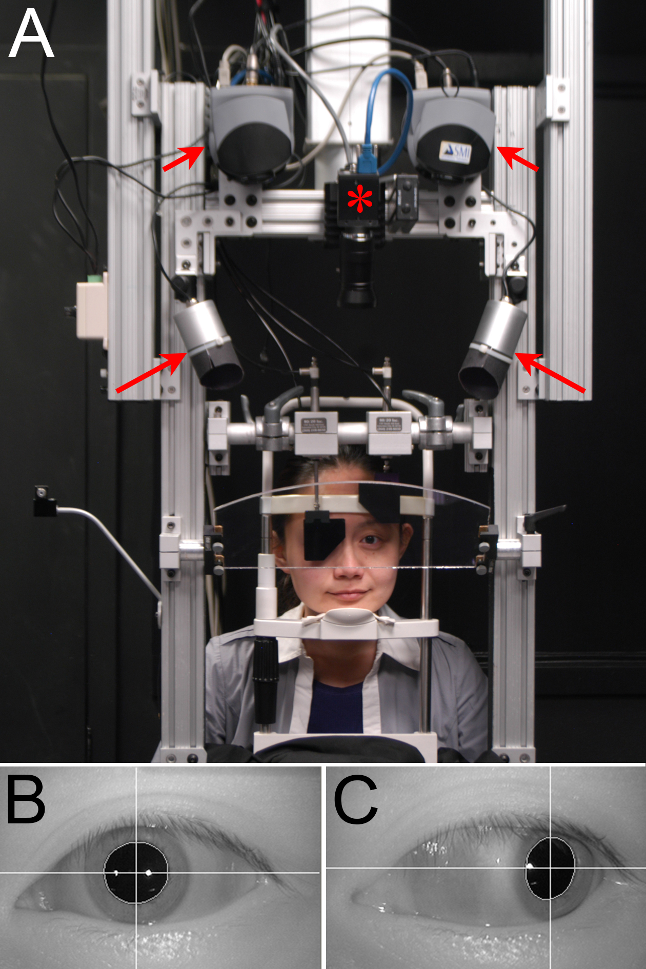
(A) Subject views tangent screen through a hot mirror, with cameras (short arrows) mounted overhead to image each eye. Infrared illuminators (long arrows) are stationed laterally. Infrared filter shutters occlude each eye without interrupting tracking. A central video camera (*) records the experiment. (B) Left eye in primary gaze, with corneal light reflexes from each illuminator. The white crosshair marks the pupil center. (C) Left eye abducted 40°, showing that the temporal light reflex in (B) remains on the cornea, so accurate tracking continues. The nasal light reflex is lost on the conjunctiva.(A) Subject views tangent screen through a hot mirror, with cameras (short arrows) mounted overhead to image each eye. Infrared illuminators (long arrows) are stationed laterally. Infrared filter shutters occlude each eye without interrupting tracking. A central video camera (*) records the experiment. (B) Left eye in primary gaze, with corneal light reflexes from each illuminator. The white crosshair marks the pupil center. (C) Left eye abducted 40°, showing that the temporal light reflex in (B) remains on the cornea, so accurate tracking continues. The nasal light reflex is lost on the conjunctiva.